Definition
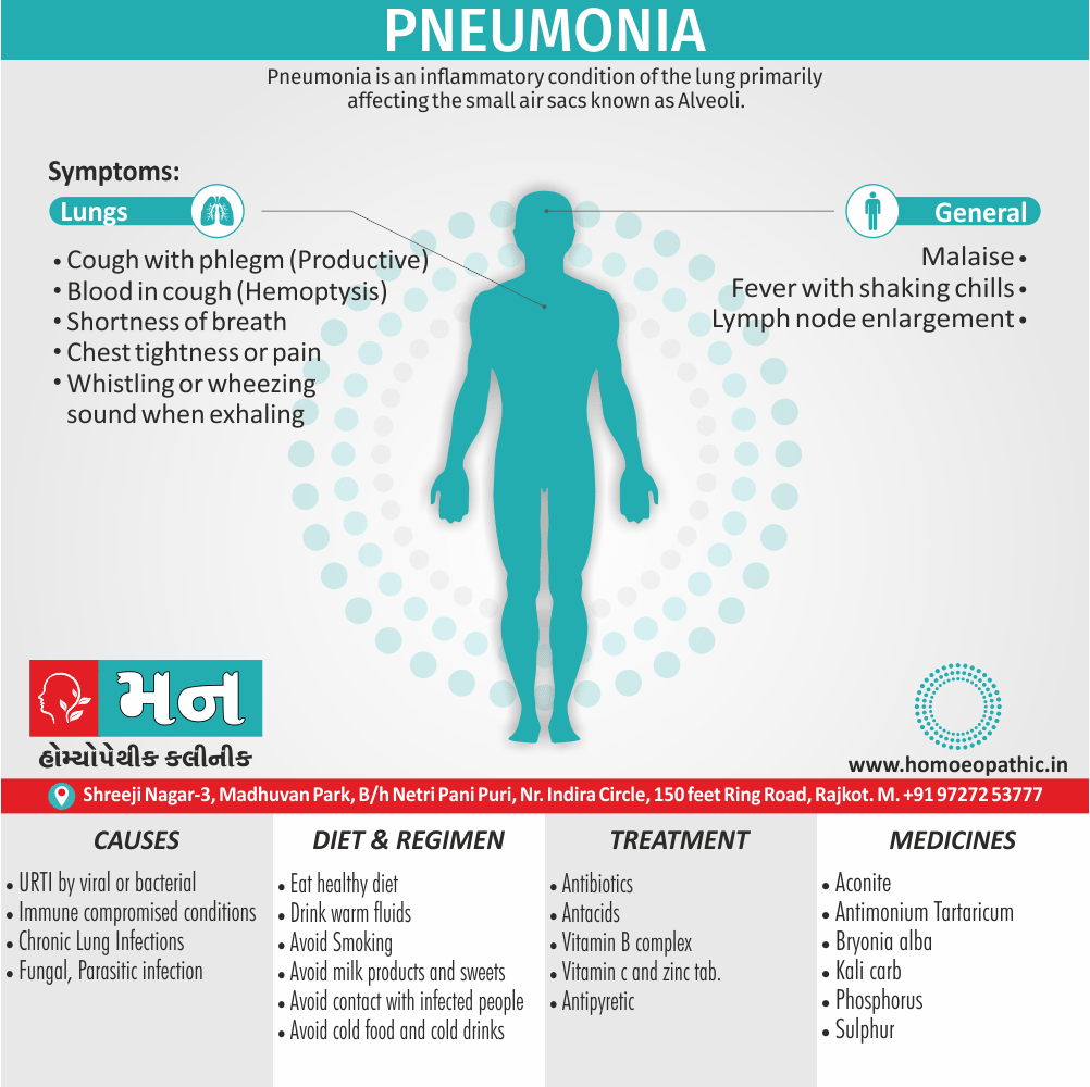
Pneumonia is an inflammatory condition of the lung primarily affecting the small air sacs known as alveoli.[1]
Overview
Epidemiology xxx
Causes
Types
Risk Factors xxx
Pathogenesis xxx
Pathophysiology
Clinical Features xxx
Sign & Symptoms
Clinical Examination xxx
Diagnosis
Differential Diagnosis
Complications xxx
Investigations
Treatment
Prevention xxx
Homeopathic Treatment
Diet & Regimen
Do’s and Dont’s xxx
Terminology xxx
References
FAQ
Also Search As xxx
Overview
Pneumonia is usually caused by infection with viruses or bacteria, and less commonly by other microorganisms. Identifying the responsible pathogen can be difficult. Diagnosis is often based on symptoms and physical examination. Chest X-rays, blood tests, and culture of the sputum may help confirm the diagnosis. The disease may be classified by where it was acquired, such as community- or hospital-acquired or healthcare-associated pneumonia.
Each year, pneumonia affects about 450 million people globally (7% of the population) and results in about 4 million deaths. With the introduction of antibiotics and vaccines in the 20th century, survival has greatly improved. Nevertheless, pneumonia remains a leading cause of death in developing countries, and also among the very old, the very young, and the chronically ill. Pneumonia often shortens the period of suffering among those already close to death and has thus been called "the old man’s friend".[1]
Epidemiology xxx
Indian epidemiology then other
Causes
This refers to the initiating factors that trigger a disease process.
- Examples of causes include:
- Pathogens: Viruses, bacteria, fungi, parasites (infectious diseases)
- Genetic mutations: Inherited or spontaneous changes in genes (genetic diseases)
- Environmental factors: Toxins, radiation, nutritional deficiencies
- Lifestyle choices: Smoking, unhealthy diet, lack of exercise (contributing factors)
[A] Predisposing factors:
Factors that predispose to pneumonia include smoking, immunodeficiency, alcoholism, chronic obstructive pulmonary disease, sickle cell disease (SCD), asthma, chronic kidney disease, liver disease, and biological aging. Additional risks in children include not being breastfed, exposure to cigarette smoke and other air pollution, malnutrition, and poverty. The use of acid-suppressing medications – such as proton-pump inhibitors or H2 blockers – is associated with an increased risk of pneumonia. Approximately 10% of people who require mechanical ventilation develop ventilator-associated pneumonia, and people with a gastric feeding tube have an increased risk of developing aspiration pneumonia. For people with certain variants of the FER gene, the risk of death is reduced in sepsis caused by pneumonia. However, for those with TLR6 variants, the risk of getting Legionnaires’ disease is increased.[2]
[B] Infection:
Bacteria
Bacteria are the most common cause of community-acquired pneumonia (in other words, CAP), with Streptococcus pneumoniae isolated in nearly 50% of cases. Other commonly isolated bacteria include Haemophilus influenzae in 20%, Chlamydophila pneumoniae in 13%, also Mycoplasma pneumoniae in 3% of cases; Staphylococcus aureus; Moraxella catarrhalis; and Legionella pneumophila. A number of drug-resistant versions of the above infections are becoming more common, including drug-resistant Streptococcus pneumoniae (in other words, DRSP) and methicillin-resistant Staphylococcus aureus (MRSA).
spreading of organisms
The spreading of organisms is facilitated by certain risk factors. Alcoholism is associated with Streptococcus pneumoniae, anaerobic organisms, also Mycobacterium tuberculosis; smoking facilitates the effects of Streptococcus pneumoniae, Haemophilus influenzae, Moraxella catarrhalis, and Legionella pneumophila. Additionally, Exposure to birds is associated with Chlamydia psittaci; farm animals with Coxiella burnetti; aspiration of stomach contents with anaerobic organisms; and cystic fibrosis with Pseudomonas aeruginosa and Staphylococcus aureus. Streptococcus pneumoniae is more common in the winter, also it should be suspected in persons aspirating a large number of anaerobic organisms.
Viruses
In adults, viruses account for about one third of pneumonia cases, and in children for about 15% of them. Commonly implicated agents include rhinoviruses, coronaviruses, influenza virus, respiratory syncytial virus (RSV), adenovirus, and parainfluenza. Herpes simplex virus rarely causes pneumonia, except in groups such as newborns, persons with cancer, transplant recipients, and people with significant burns. After organ transplantation or in otherwise immunocompromised persons, there are high rates of cytomegalovirus pneumonia.
secondary infection
Those with viral infections may be secondarily infected with the bacteria Streptococcus pneumoniae, Staphylococcus aureus, or Haemophilus influenzae, particularly when other health problems are present. Different viruses predominate at different times of the year; during flu season, for example, influenza may account for more than half of all viral cases. Outbreaks of other viruses also occur occasionally, including hantaviruses and coronaviruses. Severe acute respiratory syndrome coronavirus 2 (SARS-CoV-2) can also result in pneumonia.
Fungi
Fungal pneumonia is uncommon, but occurs more commonly in individuals with weakened immune systems due to AIDS, immunosuppressive drugs, or other medical problems. It is most often caused by Histoplasma capsulatum, Blastomyces, Cryptococcus neoformans, Pneumocystis jiroveci (pneumocystis pneumonia, or PCP), and Coccidioides immitis. Histoplasmosis is most common in the Mississippi River basin, and coccidioidomycosis is most common in the Southwestern United States. The number of cases of fungal pneumonia has been increasing in the latter half of the 20th century due to increasing travel and rates of immunosuppression in the population. For people infected with HIV/AIDS, PCP is a common opportunistic infection.
Parasites
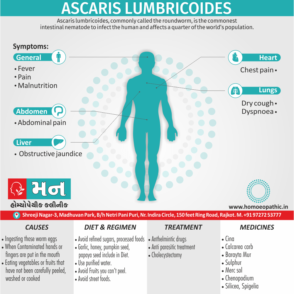
A variety of parasites can affect the lungs, including Toxoplasma gondii, Strongyloides stercoralis, Ascaris lumbricoides, and Plasmodium malariae. These organisms typically enter the body through direct contact with the skin, ingestion, or via an insect vector. Except for Paragonimus westermani, most parasites do not specifically affect the lungs but involve the lungs secondarily to other sites. Some parasites, in particular those belonging to the Ascaris and Strongyloides genera, stimulate a strong eosinophilic reaction, which may result in eosinophilic pneumonia. In other infections, such as malaria, lung involvement is due primarily to cytokine-induced systemic inflammation. In the developed world, these infections are most common in people returning from travel or in immigrants. Around the world, parasitic pneumonia is most common in the immunodeficient.
[C] Noninfectious
Idiopathic interstitial pneumonia or noninfectious pneumonia is a class of diffuse lung diseases. They include diffuse alveolar damage, organizing pneumonia, nonspecific interstitial pneumonia, lymphocytic interstitial pneumonia, desquamative interstitial pneumonia, respiratory bronchiolitis interstitial lung disease, and usual interstitial pneumonia. Lipoid pneumonia is another rare cause due to lipids entering the lung. These lipids can either be inhaled or spread to the lungs from elsewhere in the body.[1]
Types
Pneumonia is most commonly classified by where or how it was acquired:
[A]. community-acquired
[B]. Aspiration
[C]. Healthcare-associated
[D].Hospital-acquired
[E]. Ventilator-associated pneumonia.[1]
Community-acquired pneumonia [CAP]
Community-acquired pneumonia (CAP) acquire in the community, outside of health care facilities. Compared with health care–associated pneumonia, it is less likely to involve multidrug-resistant bacteria. Although the latter are no longer rare in CAP, they are still less likely.[1]
Health care–associated pneumonia (HCAP)
an infection associate with recent exposure to the health care system, including hospitals, outpatient clinics, nursing homes, dialysis centers, chemotherapy treatment, or home care. HCAP sometimes call MCAP (medical care–associated pneumonia).
People may become infected with pneumonia in a hospital; this define as pneumonia not present at the time of admission (symptoms must start at least 48 hours after admission). It likely to involve hospital-acquire infections, with higher risk of multidrug-resistant People in a hospital often have other medical conditions, which may make them more susceptible to pathogens in the hospital.
Ventilator-associated pneumonia [VAP]
occurs in people breathing with the help of mechanical ventilation. Ventilator-associated pneumonia specifically define as pneumonia that arises more than 48 to 72 hours after endotracheal intubation.[1]
It may also classify by the area of the lung affected:
[A]. Lobar pneumonia
[B]. Bronchial pneumonia
[C]. Acute interstitial pneumonia
Pneumonia in children may additionally classify based on signs and symptoms as non-severe, severe, or very severe.
The setting in which pneumonia develops is important to treatment, as it correlates to which pathogens are likely suspects, which mechanisms are likely, which antibiotics are likely to work or fail, and which complications can expect based on the person’s health status [1].
Risk Factors xxx
Risk factors are things that make you more likely to develop a disease in the first place.
Pathogenesis xxx
Pathogenesis refers to the development of a disease. It’s the story of how a disease gets started and progresses.
This is the entire journey of a disease, encompassing the cause but going beyond it.
Pathophysiology
Pathophysiology, on the other hand, focuses on the functional changes that occur in the body due to the disease. It explains how the disease disrupts normal physiological processes and how this disruption leads to the signs and symptoms we see.
Imagine a car accident. Pathogenesis would be like understanding how the accident happened – what caused it, the sequence of events (e.g., one car ran a red light, then hit another car). Pathophysiology would be like understanding the damage caused by the accident – the bent fenders, deployed airbags, and any injuries to the passengers.
In simpler terms, pathogenesis is about the "why" of a disease, while pathophysiology is about the "how" of the disease’s effects.
Pneumonia fills the lung’s alveoli with fluid, hindering oxygenation. The alveolus on the left is normal, whereas the one on the right is full of fluid from pneumonia.[1]
Mode of infection:
- Droplet infection
- Aspiration
- Blood
- Systemic
Pneumonia frequently starts as an upper respiratory tract infection that moves into the lower respiratory tract. It is a type of pneumonitis (lung inflammation). The normal flora of the upper airway give protection by competing with pathogens for nutrients. In the lower airways, reflexes of the glottis, actions of complement proteins and immunoglobulins are important for protection. Micro-aspiration of contaminated secretions can infect the lower airways and cause pneumonia. The progress of pneumonia is determined by the virulence of the organism; the amount of organism required to start an infection; and the body’s immune response against the infection.
Bacterial
Most bacteria enter the lungs via small aspirations of organisms residing in the throat or nose. Half of normal people have these small aspirations during sleep. While the throat always contains bacteria, potentially infectious ones reside there only at certain times and under certain conditions. A minority of types of bacteria such as Mycobacterium tuberculosis and Legionella pneumophila reach the lungs via contaminated airborne droplets.
Bacteria can also spread via the blood. Once in the lungs, bacteria may invade the spaces between cells and between alveoli, where the macrophages and neutrophils (defensive white blood cells) attempt to inactivate the bacteria. The neutrophils also release cytokines, causing a general activation of the immune system. This leads to the fever, chills, and fatigue common in bacterial pneumonia. The neutrophils, bacteria, and fluid from surrounding blood vessels fill the alveoli, resulting in the consolidation seen on chest X-ray.[1]
Viral
Viruses may reach the lung by a number of different routes. Respiratory syncytial virus is typically contracted when people touch contaminated objects and then they touch their eyes or nose. Other viral infections occur when contaminated airborne droplets are inhaled through the nose or mouth. Once in the upper airway, the viruses may make their way into the lungs, where they invade the cells lining the airways, alveoli, or lung parenchyma. Some viruses such as measles and herpes simplex may reach the lungs via the blood.
Lung invasion
The invasion of the lungs may lead to varying degrees of cell death. When the immune system responds to the infection, even more lung damage may occur. Primarily white blood cells, mainly mononuclear cells, generate the inflammation. As well as damaging the lungs, many viruses simultaneously affect other organs and thus disrupt other body functions. Viruses also make the body more susceptible to bacterial infections; in this way, bacterial pneumonia can occur at the same time as viral pneumonia.[1][2]
Clinical Features xxx
Tab Content
Sign & Symptoms
Sign & Symptoms
- Productive cough, fever accompanied by shaking chills, shortness of breath, sharp or stabbing chest pain during deep breaths, and an increased rate of breathing. In older people, confusion may be the most prominent sign.
- In children under five are fever, cough, and fast or difficult breathing
- Cough is frequently absent in children less than 2 months old
- More severe signs and symptoms in children may include blue-tinged skin, unwillingness to drink, convulsions, ongoing vomiting, extremes of temperature, or a decreased level of consciousness.
- Pneumonia caused by Legionella may occur with abdominal pain, diarrhea, or confusion
- It caused by Streptococcus pneumoniaeis associated with rusty colored sputum.
Other symptoms
- Pneumonia caused by Klebsiella may have bloody sputum often described as "currant jelly".
- Bloody sputum (known as hemoptysis) may also occur with tuberculosis, Gram-negative pneumonia, lung abscesses and more commonly acute bronchitis.
- Pneumonia caused by Mycoplasma pneumoniae may occur in association with swelling of the lymph nodes in the neck, joint pain, or a middle ear infection
- Viral pneumonia presents more commonly with wheezing than bacterial pneumonia.
Clinical Examination xxx
Tab Content
Diagnosis
(a). Physical examination
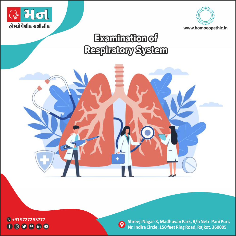
Physical examination may sometimes reveal low blood pressure, high heart rate, or low oxygen saturation. The respiratory rate may be faster than normal, and this may occur a day or two before other signs. Examination of the chest may be normal, but it may show decreased expansion on the affected side. Harsh breath sounds from the larger airways that are transmitted through the inflamed lung are termed bronchial breathing and are heard on auscultation with a stethoscope. Crackles (rales) may be heard over the affected area during inspiration. Percussion may be dulled over the affected lung, and increased, rather than decreased, vocal resonance distinguishes pneumonia from a pleural effusion.[1]
(b). Diagnosis in children
The World Health Organization has defined pneumonia in children clinically based on either a cough or difficulty breathing and a rapid respiratory rate, chest indrawing, or a decreased level of consciousness.
- A rapid respiratory rate is defined as greater than 60 breaths per minute in children under 2 months old,
- Greater than 50 breaths per minute in children 2 months to 1 year old,
- Greater than 40 breaths per minute in children 1 to 5 years old.
In children, low oxygen levels and lower chest indrawing are more sensitive than hearing chest crackles with a stethoscope or increased respiratory rate. Grunting and nasal flaring may be other useful signs in children less than five years old.
Lack of wheezing is an indicator of Mycoplasma pneumoniae in children with pneumonia, but as an indicator it is not accurate enough to decide whether or not macrolide treatment should be used.
The presence of chest pain in children with pneumonia doubles the probability of Mycoplasma pneumoniae.[1]
(c). Diagnosis in adults
In general, in adults, investigations are not needed in mild cases. There is a very low risk of pneumonia if all vital signs and auscultation are normal.
C-reactive protein (CRP)
may help support the diagnosis. For those with CRP less than 20 mg/L without convincing evidence of pneumonia, antibiotics are not recommended.
Procalcitonin
may help determine the cause and support decisions about who should receive antibiotics. Antibiotics are encouraged
if the procalcitonin level reaches 0.25 μg/L, strongly encouraged
it reaches 0.5 μg/L, and strongly discouraged
if the level is below 0.10 μg/L.
In people requiring hospitalization,
- Pulse oximetry,
- Chest radiography
- Blood tests– including a complete blood count, serum electrolytes,
- C-reactive protein level, and possibly
- Liver function tests– are recommended.
The diagnosis of influenza-like illness can be made based on the signs and symptoms; however, confirmation of an influenza infection requires testing. Thus, treatment is frequently based on the presence of influenza in the community or a rapid influenza test.[1]
Differential Diagnosis
Several diseases can present with similar signs and symptoms to pneumonia, such as:
- Chronic obstructive pulmonary disease,
- Asthma,
- Pulmonary edema,
- Bronchiectasis,
- Lung cancer, and
- Pulmonary emboli.[2]
Complications xxx
Complications are what happen after you have a disease. They are the negative consequences of the disease process.
Investigations
Other Investigation
- X-Ray chest
- CT-Scan
- Lung ultrasound
Treatment
Antibiotics by mouth, rest, simple analgesics, and fluids usually suffice for complete resolution.
However, those with other medical conditions, the older people, or those with significant trouble breathing may require more advanced care. If the symptoms worsen, the pneumonia does not improve with home treatment, or complications occur, hospitalization may require.
Bacterial
- Antibiotics improve outcomes in those with bacterial pneumonia. The first dose of antibiotics should give as soon as possible. Increased use of antibiotics, however, may lead to the development of antimicrobial resistant strains of bacteria.
- Antibiotic use also associate with side effects such as nausea, diarrhea, dizziness, taste distortion, or headaches
Worldwide spread
- In the UK, treatment before culture results with results with amoxicillin recommend as the first line for community-acquired pneumonia, with doxycycline or clarithromycin as alternatives.
- In North America, amoxicillin, doxycycline, and in some areas a macrolides(such as azithromycin or erythromycin) is the first-line outpatient treatment in adults.
- In children with mild or moderate symptoms, amoxicillin taken by mouth is the first line.
- The use of fluoroquinolones in uncomplicated cases discourage due to concerns about side-effects and generating resistance in light of there being no greater benefit.
- Recommendations for hospital-acquired pneumonia include third- and fourth-generation cephalosporins, carbapenems, fluoroquinolones, aminoglycosides, and vancomycin.
Viral
- Neuraminidase inhibitors may use to treat viral pneumonia caused by influenza viruses (influenza A and influenza B).[1]
- No specific antiviral medications recommend for other types of community acquired viral pneumonias including SARS coronavirus, adenovirus, hantavirus, and parainfluenza virus.
- Influenza A may treat with rimantadine or amantadine, while influenza A or B may treat with oseltamivir, zanamivir or peramivir.
- These are of most benefit if they start within 48 hours of the onset of symptoms. Many strains of H5N1 influenza A, also known as avian influenza or "bird flu", have shown resistance to rimantadine and amantadine. The use of antibiotics in viral pneumonia recommend by some experts, as it is impossible to rule out a complicating bacterial infection. The British Thoracic Society recommends that antibiotics withheld in those with mild disease. The use of corticosteroids is controversial.
Aspiration
- In general, aspiration pneumonitis treat conservatively with antibiotics indicated only for aspiration pneumonia. The choice of antibiotic will depend on several factors, including the suspected causative organism and whether pneumonia acquire in the community or developed in a hospital setting. Common options include clindamycin, a combination of a beta-lactam antibiotic and metronidazole, or an aminoglycoside. Corticosteroids sometimes use in aspiration pneumonia, but there is limited evidence to support their effectiveness.[1]
Prevention xxx
Tab Content
Homeopathic Treatment
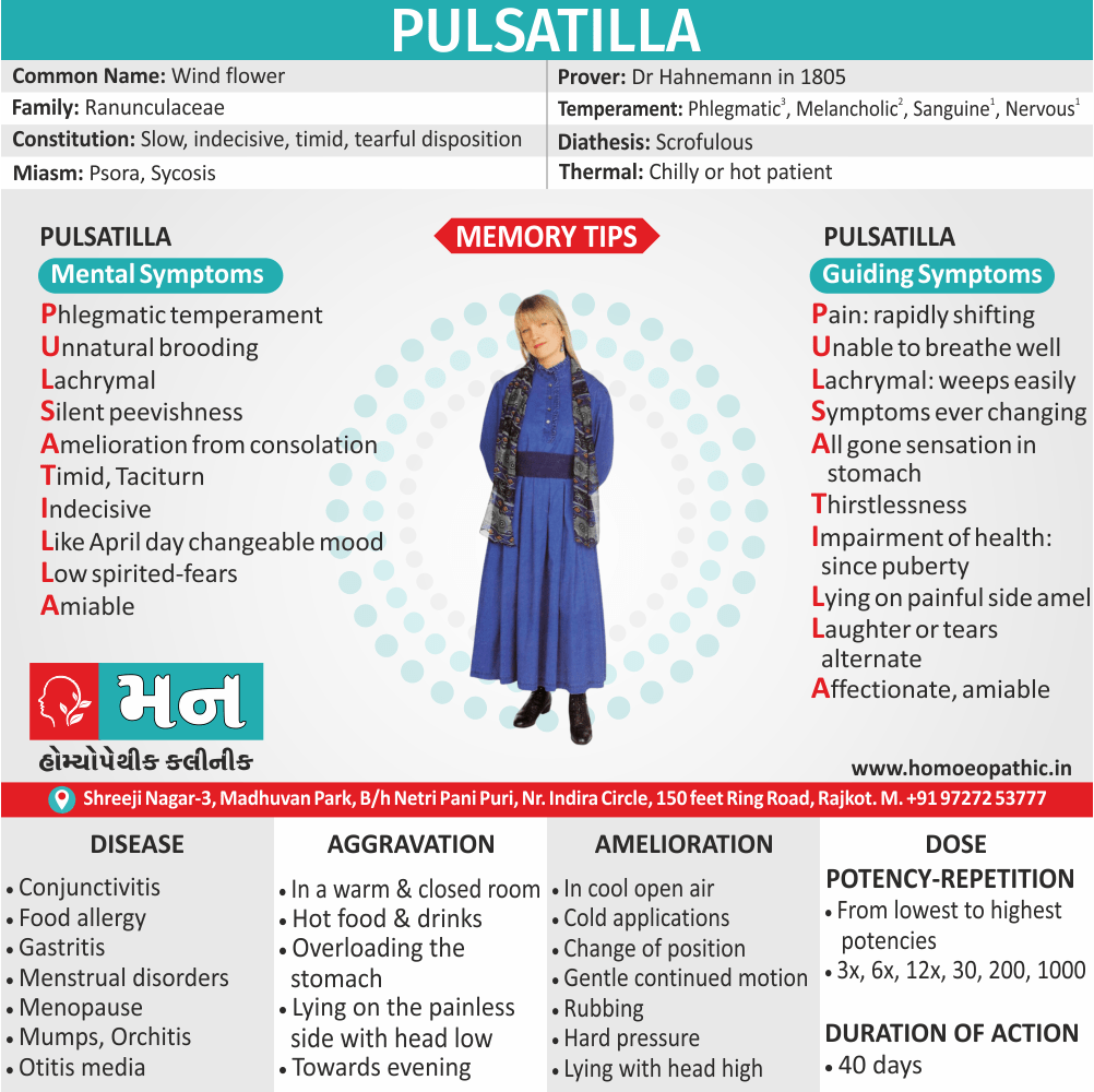
Homeopathy a form of alternative medicine used for the treatment of pneumonia, is based on the theory of individualization. It takes into account the lifestyle, hereditary factors, personality as well as the medical history of a person & aims to treat the person as a whole including their psychological & physiological health.
Some of the homeopathic remedies that are effective against pneumonia are [4],
Ferrum phosphoricum
This, like Aconite, is a remedy for the first stage before exudation takes place, and, like Aconite, if there be any expectoration it is thin, watery and blood streaked. It is a useful remedy for violent congestions of the lungs, whether appearing at the onset of the diseases or during its course, which would show that the inflammatory action was extending; it thus corresponds to what term secondary pneumonias, especially in the age and debilitated. There high fever, oppress and hurry breathing, and bloody expectoration, very little thirst; there are extensive rales, and perhaps less of that extreme restlessness and anxiety that characterizes Aconite. This remedy, with kali muriaticum, forms the Schuesslerian treatment of this disease.
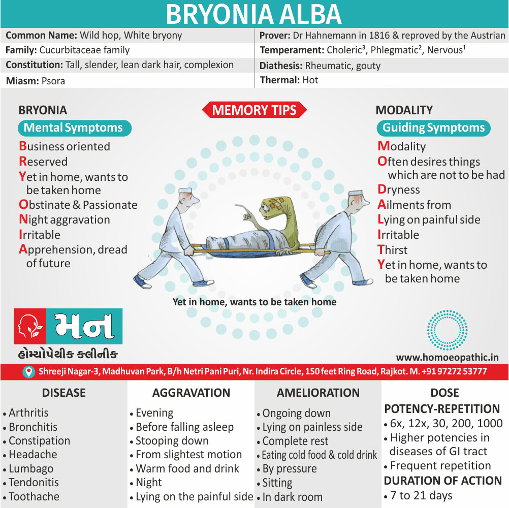
Bryonia Alba
is the remedy for pneumonia; it furnishes a better pathological picture of the disease than any other, and it comes in after Bryonia is looser and moister than that of Aconite, and there are usually sharp stitching pleuritic pains, the cough of Bryonia is also hard and dry at times and the sputum is scanty and rust colored, so typical of pneumonia. There may circumscribe redness of the cheeks, slight delirium and apathy; the tongue will most likely dry, and the patient will most likely dry, l and the patient will want to keep perfectly quiet. It a right-side remedy and attacks the parenchyma of the lung, and perhaps more strongly indicate in the croupous form of pneumonia.
The patient dreads to cough and holds his breath to prevent it on account of the pain it causes; it seems as though the chest walls would fly to pieces. The pains in the chest, besides being worse by motion and breathing, are relieved by lying on the right or painful side, because this lessens the motions; of that side. Coughs which hurt distant parts of the body call for Bryonia. Phosphorus most commonly follows Bryonia in pneumonia, and is complementary. In pneumonias complicated by pleurisy Bryonia is the remedy, par excellence. Halbert believes that Cantharis relieves the painful features of the early development of the exudate better than any other remedy, a hint which comes from Dr.Jousset, who used the remedy extensively.[3]
Kali muriaticum
Since the advent of Schussler’s this has been a favorite remedy with some physicians, and not without a good ground for its favoritism. Clinical experience has proved that this drug in alternation with Ferrum Phosphoricum constitutes a treatment of pneumonia which has been very successful in many hands. The symptoms calling for Kali muriaticum as laid down by Schussler are very meager, it given simply because there is a fibrinous exudation in the lung substance. There is a white, viscid expectoration and the tongue coated white. It better suited to the second stage, for when the third stage appears with its thick, yellowish expectoration it replace by Kali Sulphuricum in the biochemic nomenclature.[3]
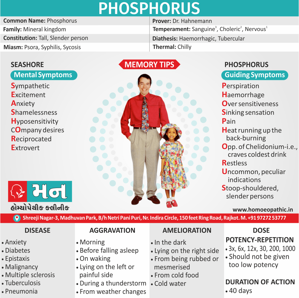
Phosphorus
is "the great mogul of lobar pneumonia." It should remembered that Phosphorus is not, like Bryonia, the remedy when the lungs completely hepatized, although it is one of the few drugs which have known to produce hepatization. When bronchial symptoms are present it is the remedy, and cerebral symptoms during pneumonia often yield better to Phosphorus than to Belladonna. There is cough; with pain under sternum, as if something torn loose; there is pressure across the upper part of the chest and constriction of the larynx; there pressure across the upper part of the chest and constriction of the larynx; there are mucous rales, labored breathing, sputa yellowish mucus, with blood streaks therein, or rust colored, as under Bryonia.
Relationship of Phosphorus
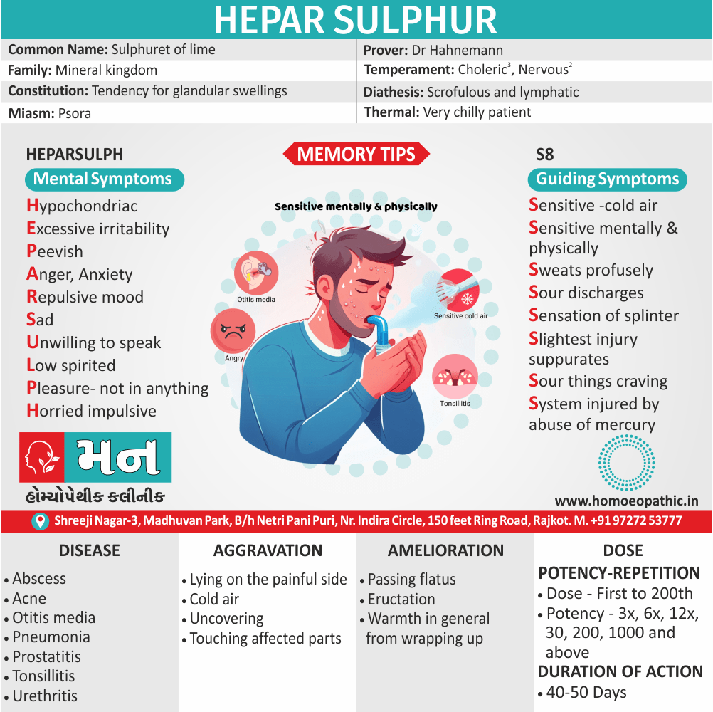
After Phosphorus, Hepar Sulphur. naturally follows as the exudate begins to often; it is the remedy of the third stage, the fever is; of a low character. Tuberculinum. In lobular pneumonia this remedy surpasses Phosphorus or Antimonium tartaricum, and competent observers convinced that it has an important place in the treatment of pneumonia; some using it in very case intercurrently; doses varying from 6x to 30x. When typhoid symptoms occur in the course of pneumonia then Phosphorus will come in beautifully.
Phosphorus follows Bryonia well, being complementary to it. There is also a sensation as if the chest were full of blood, which causes an oppression; of breathing, a symptom met with commonly enough in pneumonia. Hughes maintains that Phosphorus should give in preference to almost any medicine in acute chest affections in young children. Lilienthal says Phosphorus is our great tonic to the heart and lungs. Hyoscyamus. Dr. Nash considers this remedy one; of the best in typhoid pneumonia, meaning that it is more frequently indicated than any other.[3]
Chelidonium
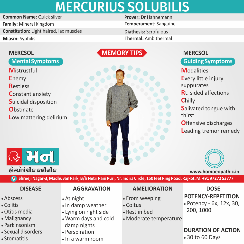
Bilious pneumonia is, perhaps more often indicative of Chelidonium than of any other remedy. there are stitching pains under the right scapula, loose rattling cough and difficult expectoration, oppression; of chest, as under Antimonium tartaricum, and fan-like motions of the alae nasi, as under Lycopodium. Mercurius is quite similar in bilious pneumonia; the stools will decide, those of Mercurius being slimy and accompan by tenesmus; the expectoration is also apt to blood-streak. With Chelidonium there is an excess of secretion in the tubes, which; is similar to Antimonium tartaricum, and an inability to raise the same. It has greatly praise in catarrhal pneumonia of young children where there is plentiful secretion and inability to raise it. The right lung more often affect in cases calling for Chelidonium.[3]
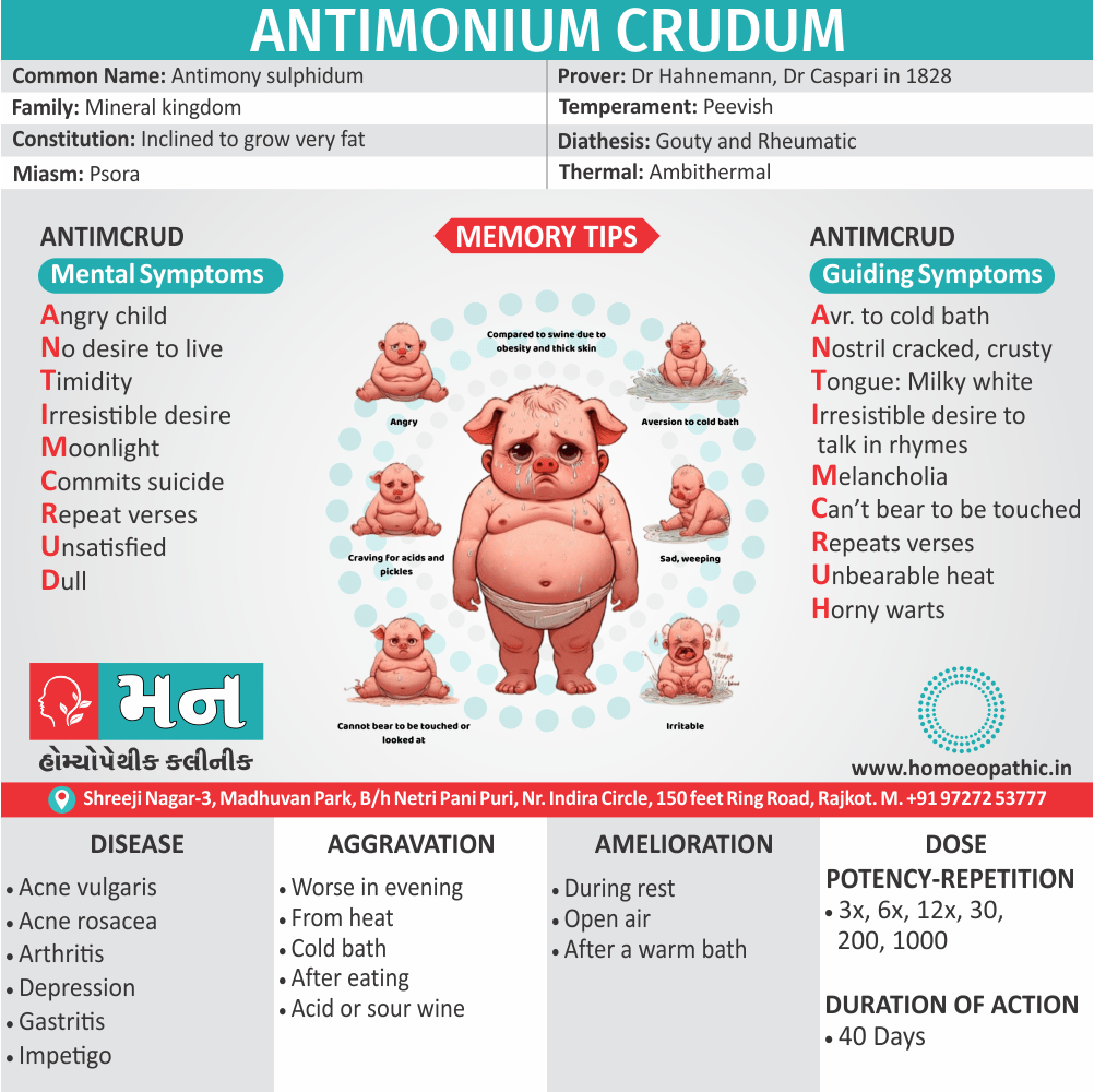
Antimonium tartaricum
This drug especially indicated in pneumonia and pleuro-pneumonia at the stage of resolution. There are fine moist rales heard all over the hepatized portion of the lungs; these are different from the Ipecac rales; they are fine, while those of Ipecac are coarse. With Antimonium tartaricum there is great oppression of breathing, worse towards morning, compelling the patient to sit up to breath. There are also sharp, stitching pains and high fever, as under Bryonia, and it, perhaps, more closely corresponds to the catarrhal form than it does to the croupous. Bilious symptoms, if present, do not contra-indicate, as there are many of these in its pathogenesis. There is one peculiar symptom, the patient feels sure that the next cough will raise the mucus, but it does not. When there is deficient reaction, as in the aged or; in very young children, this remedy particularly indicated.
Kali carbonicum
is, perhaps, more similar to Bryonia than any drug in the symptom of sharp, stitching pains in the chest. These are worse by motion, but, unlike Bryonia they come whether the patient moves or not, and are more in the lower part of the right lung. In pneumonia with intense dyspnoea and a great deal of mucus on the chest, which, like in all of the Kalis, raise with difficulty, wheezing and whistling breathing, Kali carbonicum is the remedy, especially if the cough tormenting. It comes in with benefit ofttimes where Antimonium tartaricum and Ipecac have failed to raise the expectoration. Kali bichromicum may indicate by its well-known tough, stringy expectoration.[3]
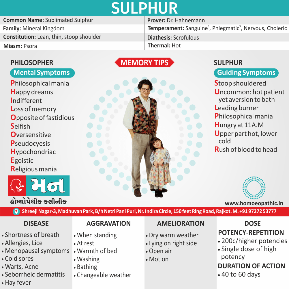
Sulphur
A remedy to use in any stage of pneumonia. It will prevent, if given in the beginning, if the symptoms indicate it. It will prevent hepatization and cause imperfect and slow resolution to react. When the case has a typhoid tendency and the lung and the lung tends to break down, where there are rales, muco-purulent expectoration slow speech, dry tongue and symptoms of hectic, Sulphur is the remedy. Weakness and faintness are characteristic symptoms. Dr. G. J. Jones says a dyspnoea occurring at night between 12 and 2 causing the patient to sit up in bed is a valuable symptom.
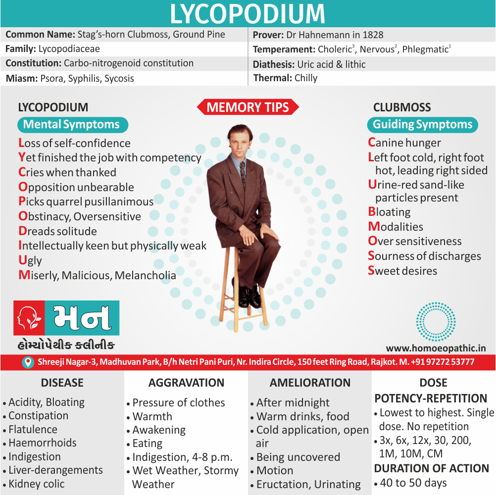
Its field especially in neglect pneumonias in psoric constitutions, with tendency to develop into tuberculosis. In purulent expectoration Sanguinaria is the better remedy, especially where it is offensive even to the patient himself. If the lung hepatize, the patient at night restless and feverish, ulceration threatened, and there is no tendency to recuperation then one may depend upon Sulphur. Lycopodium also; a most useful remedy in delay or partial resolution. There is a tightness across the chest, aching over lungs, general weakness. Hughes says it is the best remedy where the case threatens to run into acute phthisis.[3]
Diet & Regimen
Diet & Nutrition
- Drink plenty of fluids and fresh juice of fruits and vegetables.
- Consume hot vegetable soups – tomato, sweet corn etc.
- Add a pinch of turmeric to your diet as it has anti-inflammatory properties.
- Initially diet should consist of fresh fruits and vegetables, when better can start with grains and little of protein in diet.
- Milk and milk products and sweets should be avoided to decrease mucus production.
- Onion and garlic should be consumed it is beneficial.
- Boil a mixture of Bishops weed (Ajwain), tea leaves and water and inhale the steam, helps to decongest. Do this at least 2-3 times a day.
- Gargle with warm water, a pinch of salt and turmeric to sooth your throat.
- Consume lots of vitamin A, maintains the integrity of the respiratory mucosa: Liver oils of fish like cod, shark, and halibut are richest source of vitamin A.
Other dietary measures
- Egg, milk and milk products, meat, fish, kidney and liver.
- Yellow orange-colored fruits and vegetables, dark green leafy vegetables.
- Increase intake of vitamin C, it has antioxidant property: foods of animal origin are poor in vitamin C.
- Citrus fruits, green vegetables.
- Include zinc in your diet, it boosts up your immunity:
- Meat, poultry and milk, sea food – shell fish, crab, shrimp, and sea plants etc.
- Plant foods are low in zinc, Whole wheat grains provide good amount of zinc.
- Avoid sharing things such as towels, beverages with infected people.
- Avoid contact with infected people.
- Avoid crowded places.
- Avoid smoking and alcohol.[5]
Do’s and Dont’s xxx
Tab Content
Terminology xxx
Tab Content
References
FAQ
Frequently Asked Questions
What is Pneumonia?
Pneumonia is an inflammatory condition of the lung primarily affecting the small air sacs known as alveoli.
Homeopathic Medicines used by Homeopathic Doctors in treatment of Pneumonia?
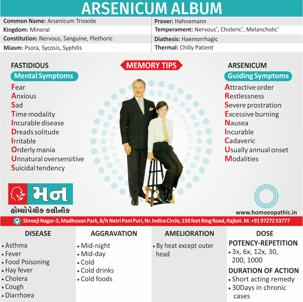
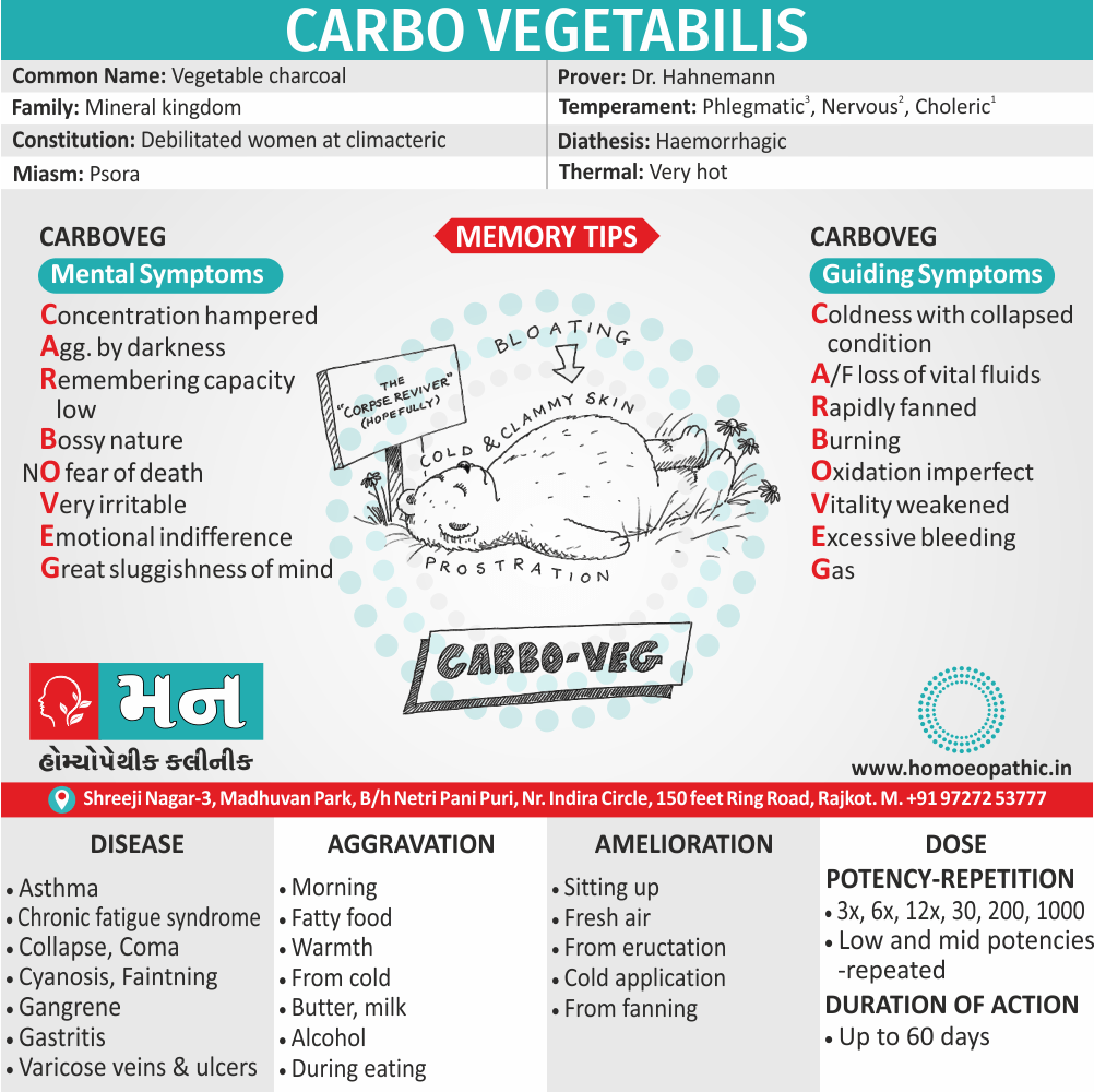
- Bryonia Alba
- Kali Muriaticum
- Phosphorus
- Chelidonium
- Antimonium tartaricum
- Kali carbonicum
- Sulphur
What causes Pneumonia?
- Smoking, immunodeficiency, alcoholism
- Pollution, malnutrition, and poverty
- Bacteria, Viruses, Fungi, Parasites
- Diffuse alveolar damage
- Organizing pneumonia
- Nonspecific interstitial pneumonia
- Lymphocytic interstitial pneumonia
What are the symptoms of Pneumonia?
- Productive cough
- Fever accompanied by shaking chills
- Shortness of breath
- Sharp or stabbing chest pain during deep breaths
- Increased rate of breathing
- Blue-tinged skin
- Unwillingness to drink
- Convulsions
- Ongoing vomiting
- Extremes of temperature
- Decreased level of consciousness
Give the types of Pneumonia?
- Community-acquired
- Aspiration
- Healthcare-associated
- Hospital-acquired
- Ventilator-associated pneumonia
Also Search As xxx
Frequently Asked Questions (FAQ)
XYZ
XXX
XYZ
XXX
XYZ
XXX
How can I find reputable homeopathy clinics or homeopathic doctors in my area?
You can found Homeopathic Clinic For XXXX by searching for
Specific city Examples are
You can also search for near you Examples are
