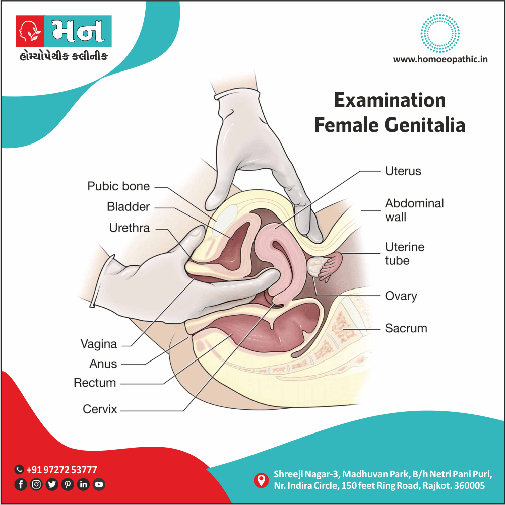
Examination Of The Female Genitalia
Definition
Examination of female genitalia, also known as a pelvic exam, is a crucial part of women’s health care. It aims to assess the health of the reproductive organs and detect any potential abnormalities or diseases.
The examination of female genitalia preced by an in-depth history taking, including present complaints, past and personal history (previous medical, gynaecological and surgical illnesses and interventions), family history, marital and sexual history (when relevant), menstrual history and obstetric history (when relevant).
It must include a thorough general and systemic examination, taking into consideration the parameters of built (e.g. obese/thin), nutrition, pallor, jaundice, enlarged lymph nodes, swellings in the neck, oedema of feet, pulse, and blood pressure. The cardiovascular and respiratory systems must also cursorily examine.
There isn’t a perfect synonym for "examination of female genitalia" because it’s a specific medical term. However, depending on the context, you could use these terms that capture the main idea:
- Pelvic exam:
This is the most common and appropriate term used by medical professionals for an examination of the female genitalia. It includes examining the external genitalia, vagina, cervix, uterus, and ovaries.
- Genital exam:
This is a broader term that could encompass an examination of both male and female genitalia.
Internal exam:
This term refers to any examination that involves inserting instruments or fingers into the body, and it could be used in the context of a pelvic exam.
It’s important to use the correct terminology depending on the situation. If you’re talking to a doctor, "pelvic exam" is the best choice. If you’re talking in a more general setting, "examination of female genitalia" might be appropriate, but "pelvic exam" would still be more precise.
BREAST EXAMINATION
PELVIC EXAMINATION
External Examination
Internal Examination:
Additional Tests
Patient Comfort and Privacy
Terminology
BREAST EXAMINATION
BREAST EXAMINATION IN EXAMINATION OF THE FEMALE GENITALIA:
A routine breast examination must done especially above the age of 30 years, to detect any abnormality or pathology.
ABDOMINAL EXAMINATION IN EXAMINATION OF THE FEMALE GENITALIA:
The patient should first instruct to empty the urinary bladder before coming in for examination. Examination done with patient lying flat on the table with thighs slightly flexed and abducted.
INSPECTION:
The abdomen observe for the presence of umbilical eversion (e.g. large tumours, ascites, pregnancy), straie (e.g. pregnancy, obesity and parous women), surgical scars especially in the lower abdomen also prominent veins. Asking the patient to cough can elicit the presence of an incisional hernia. Besides this, Movements of abdomen are observed, especially lower abdomen, to look for pelvic peritonitis.
PALPATION:
The abdomen palpate for any swellings arising from the pelvis. The consistency, mobility and margins are to felt. In case of pelvic tumours, the lower border of the swelling cannot felt, whereas ovarian neoplasms can palpate below the lower pole. Testing for fluid thrill will elicit if the swelling is cystic or not. Intense tenderness on palpating below the umbilicus is suggestive of pelvic peritoneal irritation which can indicate PID, ectopic pregnancy, red degeneration of fibroid or twist ovarian cyst.
PERCUSSION in Examination of Female Genitalia :
Pelvic tumours are dull to percussion but the flanks are resonant. In cases of ascites, the flanks will dull to percussion and shifting dullness can elicit. (Ascites may associate with Pseudo-Meig’s syndrome, malignancy and tuberculous peritonitis).
AUSCULTATION in Examination of Female Genitalia:
Auscultation done to affirm the presence of bowel sounds. Absence of bowel sounds seen in cases of generalize peritonitis. Auscultation also reveals uterine soufflé in vascular fibroids and pregnant uterus and the presence of fetal heart sounds in pregnancy.
PELVIC EXAMINATION
PELVIC EXAMINATION IN EXAMINATION OF THE FEMALE GENITALIA:
EXTERNAL GENITAL EXAMINATION:
The distribution of pubic hair note, as well as whether any anatomic abnormalities are present in the clitoris, labia and perineum. Presence of any vaginal discharge or blood is looked for and noted. The patient ask to cough/strain to elicit the presence of a genital prolapse or stress urinary incontinence. Labia separate using fingers and the external urethral meatus also character of hymen are noted. Usually, the openings of the Bartholin’s ducts cannot be visualized, but if they can seen, an inflammation of the ducts suspect.
VAGINAL EXAMINATION:
SPECULUM EXAMINATION
Speculum examination ideally done prior to a bimanual examination especially when a Pap smear needs to take or a specimen of any discharge needs to collect for bacteriological investigations. Also, if a lesion is present in the cervix, it may bleed on digital examination, making it difficult to visualize during a subsequent speculum examination.
The Cusco’s self-retaining speculum use for satisfactory inspection of the cervix and vaginal fornices and for collection of cervical and vaginal smears. The anterior vaginal wall, however, best visualize with a Sims’ speculum. It permits the evaluation of presence of cystocele or rectocele.
DIGITAL EXAMINATION
In brief, Digital examination always carried out using well-lubricated, gloved fingers.
Cervix in Examination of Female Genitalia
Two fingers of the right hand introduce in the vaginal introitus and moved up to the fornices. Whether the anterior or posterior lip of cervix felt first is noted. If the anterior lip felt first, it means that the cervix push downwards and hence the uterus anteverted. If the posterior lip felt first, the cervix upwards and the uterus retrovert. After making note of the position of cervix, its consistency note; in the pregnant state, the cervix feels soft, whereas in the non-pregnant state, it feels as firm as the tip of the nose. Movement of the cervix elicits pain in cases of ectopic pregnancy and acute salpingo-oophoritis .
Uterus
A bimanual examination done by placing the left hand over the hypogastrium. The fingers in the introitus are then used to push the fornices up so that the uterus (if anteverted) then palpated between both hands and its shape, size, tenderness and any pathology is noted. A retroverted uterus can examine using the fingers present internally only, through the posterior fornix.
Adnexa in Examination of Female Genitalia
Lateral fornices then felt and push up so that any swellings in the lateral part of the pelvis can palpate bimaually. A mass felt separate from the uterus (or a mass that does not move on movement of the cervix) indicative of an adnexal swelling, the most common of which are ovarian cyst, paraovarian cyst, ovarian tumour, chronic ectopic pregnancy or tubo-ovarian mass.
Fallopian tubes are non-palpable, unless they enlarge. Ovaries usually are non-palpable.
Pouch of Douglas
Generally, The pouch of Douglas palpate from the posterior fornix and swellings, if any, note. Additionally, Swelling felt in this area can indicated a retroverted uterus, loaded rectum, pelvic inflammatory masses, chocolate cyst of ovary, pelvic abscess or ovarian neoplasm.
RECTAL EXAMINATION in Examination of Female Genitalia
A rectal examination can do additionally to a vaginal examination or on its own. The lower bowel should ideally empty during a rectal examination and it always carry out with well-lubricated glove fingers.
A rectal examination carried out as part of a gynaecological work-up in cases of i.e.:
- Virgins also children
- Carcinoma of cervix to determine the extent of its spread posteriorly
- Pelvic endometriosis
- Vaginal atresia
- Rectocele to distinguish it from an enterocele
- Swelling in the pouch of Douglas (already determined by vaginal examination), for better appreciation
External Examination
Internal Examination:
A speculum, a duck-billed instrument, is gently inserted into the vagina to visualize the cervix and vaginal walls. This allows for the collection of cells for a Pap smear, which screens for cervical cancer.
Bimanual Examination:
The healthcare provider inserts two lubricated, gloved fingers into the vagina while gently pressing on the lower abdomen with the other hand. This helps assess the size, shape, and position of the uterus and ovaries, and detect any tenderness or masses.
Additional Tests
Additional Tests:
Depending on the patient’s medical history and symptoms, additional tests may be performed during the pelvic exam. These could include:
- Rectovaginal Examination: The healthcare provider inserts one finger into the vagina and another into the rectum to assess the muscles and tissues between the vagina and rectum.
- STI Testing: Samples may be collected to test for sexually transmitted infections (STIs).
- Urine Test: A urine sample may be collected to check for infections or other conditions.
Patient Comfort and Privacy
Patient Comfort and Privacy:
Maintaining patient comfort and privacy is paramount during the pelvic exam. The healthcare provider should explain each step of the procedure, address any concerns, and ensure the patient feels relaxed and comfortable throughout the process.
Terminology
Terminology
The examination of female genitalia involves a specialized vocabulary used to describe the anatomy and any potential findings. Here are some common terminologies used in such articles:
External Genitalia:
- Vulva: The external female genitals, including the mons pubis, labia majora, labia minora, clitoris, and vaginal opening.
- Mons Pubis: The rounded fatty pad of tissue covering the pubic bone.
- Labia Majora: The outer, larger folds of skin surrounding the vulva.
- Labia Minora: The inner, smaller folds of skin within the labia majora.
- Clitoris: The highly sensitive organ at the top of the vulva responsible for sexual pleasure.
- Vestibule: The area enclosed by the labia minora, containing the urethral opening and vaginal opening.
- Perineum: The area between the vaginal opening and the anus.
Internal Genitalia:
- Vagina: The muscular canal connecting the external genitals to the cervix and uterus.
- Cervix: The lower, narrow end of the uterus that opens into the vagina.
- Uterus: The pear-shaped organ where a fetus develops during pregnancy.
- Fallopian Tubes: The tubes that connect the ovaries to the uterus and where fertilization typically occurs.
- Ovaries: The almond-shaped organs that produce eggs and hormones.
Other Relevant Terms:
- Speculum: An instrument used to open the vagina during examination.
- Bimanual Exam: An examination where the healthcare provider inserts fingers into the vagina and presses on the abdomen to feel the internal organs.
- Pap Smear: A screening test for cervical cancer.
- Colposcopy: A procedure to closely examine the cervix, vagina, and vulva for abnormalities.
Frequently Asked Questions (FAQ)
What is Examination Of The Female Genitalia?
Definition
The examination of female genitalia preced by an in-depth history taking, including present complaints, past and personal history (previous medical, gynaecological and surgical illnesses and interventions), family history, marital and sexual history (when relevant), menstrual history and obstetric history (when relevant).
What is included in Examination Of The Female Genitalia?
Examination of the female genitalia
- Breast examination
- Abdominal Examination
- Pelvic Examination
How to examine abdomen in Examination Of The Female Genitalia?
In 4 steps:
- Inspection
- Palpation
- Percussion
- Auscultation
What are the methods of Vaginal Examination?
Method
- Speculum Examination
- Digital Examination
What is the purpose of a female genital examination?
Purpose
A female genital examination is a routine medical procedure to assess the health of the external and internal reproductive organs. It helps detect early signs of infections, sexually transmitted diseases, cancers, or other abnormalities.
