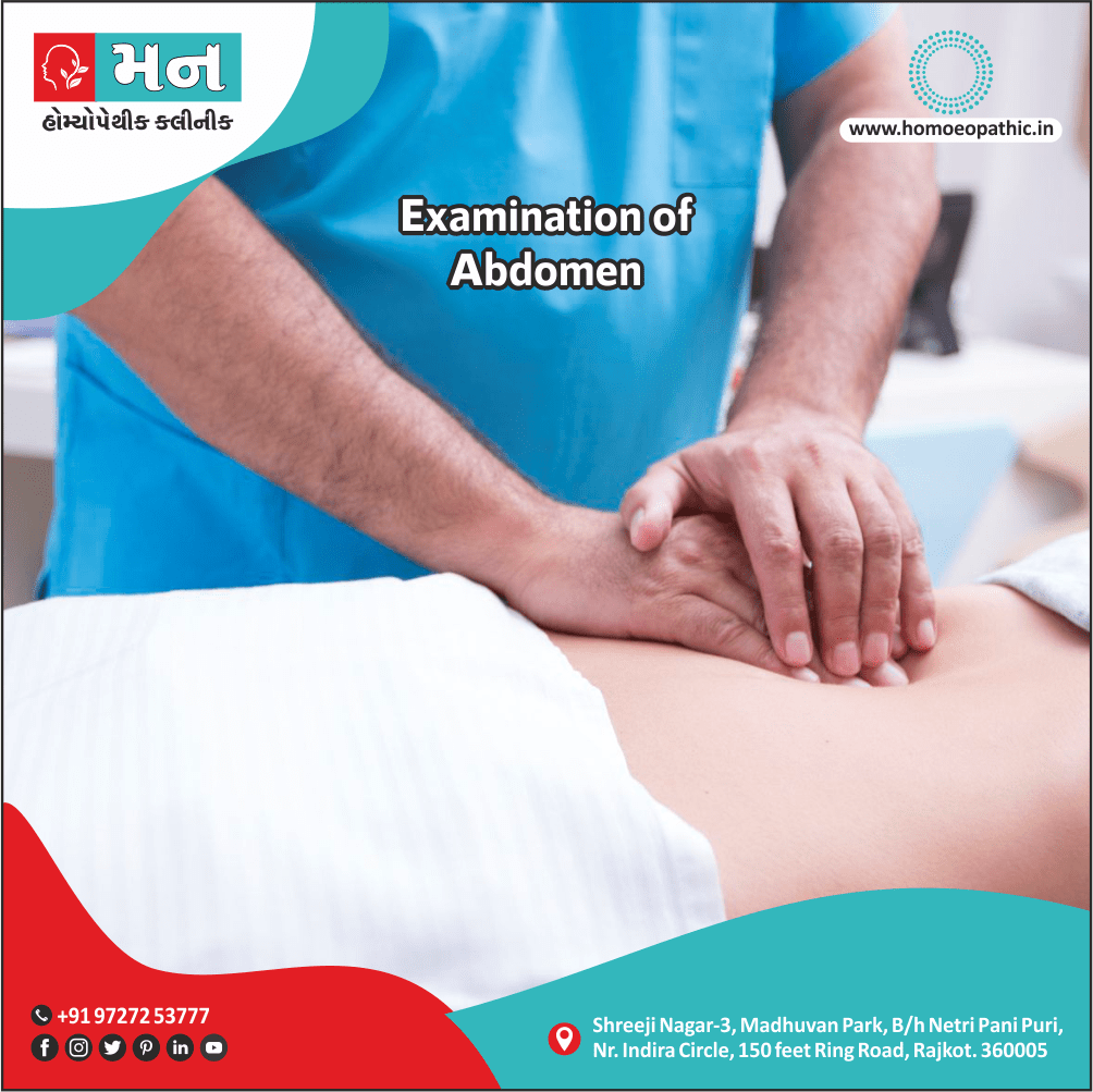
Abdominal Examination
Definition
Abdominal Examination consider Symptoms of GI or Abdominal diseases are often vague, and signs of abnormality few unless the disease is advanced. Assessment of the nutrition status is particularly very important for coming to a diagnosis, in GI diseases or digestive problems. A proper history, along with a thorough abdominal examination, aids the diagnostic process. So, if you have abdominal pain or digestive problems, consult a homeopathic doctor.
While you can use the term "abdominal examination" to refer to the entire process, you can also use more specific terms.
"Abdominal assessment" is a broader term encompassing the entire evaluation. However, if you want to focus on specific aspects, you can use terms like "inspection" and "palpation."
Inspection involves visually examining the abdomen. In contrast, palpation involves using your hands to feel the abdomen and assess for tenderness, masses, or other abnormalities. And of course, "belly check" is a more informal term for a general examination.
You can also use more general terms for examination, such as:
- Abdominal checkup
- Abdominal evaluation
- Physical examination of the abdomen
INSPECTION
PALPATION
PERCUSSION
AUSCULTATION
INSPECTION
INSPECTION
To begin the abdominal examination, position the patient supine on the examination table. After ensuring the patient is comfortable, stand on their right side and inspect the abdomen under good lighting. During inspection, visually scan the area from the xiphisternum to the symphysis pubis.
SHAPE:
- The normal shape of the abdomen is scaphoid or boat-shaped. Abdominal Distention, however, may be generalized or localized. Generalized distention may be due to fat, fluid, flatus, feces, or pregnancy. Conversely, localized distention may be seen in small bowel obstruction, gross enlargement of the spleen, liver, or ovaries.
- After examining the abdomen, the doctor will shift their focus to the groin area. Here, a meticulous Abdominal examination is crucial, particularly to identify potential inguinal or femoral hernias. If no visible swelling is present at rest, the doctor will then ask the patient to cooperate with a specific maneuver. They will instruct the patient to turn onto their side and cough. This action can sometimes cause a hernia, if present, to become visible. However, to definitively differentiate between an inguinal and femoral hernia, Abdominal palpation is the next essential step.
SKIN:
- Look for any form of pigmentation on the abdominal wall. For example, linea nigra, a dark line below the umbilicus, indicates pregnancy. Similarly, erythema ab igne, a brownish pigmentation caused by constant heat application, suggests long-standing pain, such as in chronic pancreatitis.
- In Abdominal Examination Any cause of distention may result in smooth and glossy skin, whereas the resolution of previous distention may cause wrinkled skin.
- Abdominal Examination striae, commonly known as stretch marks, result from the rupture of subepidermal connective tissue due to abdominal distention, whether present or past. They commonly appear after pregnancy.
- Dilated veins indicate venous obstruction of any form. On the abdominal wall, IVC or portal vein obstruction usually causes them.
- Spider nevi commonly occur in alcohol use disorder, cirrhosis, pregnancy, rheumatoid arthritis, and thyrotoxicosis. However, they sometimes appear in normal individuals as well. A spider nevus has a central arteriole with radiating small vessels. They pulsate and blanch with pressure.
- Finally, look for any scars, old or recent, as they may indicate a previous injury or surgery.
UMBILICUS:
Abdominal Examination ,The umbilicus is normally slightly inverted and retracted. However, in ascites, it acquires a smiling face (transverse stretch) or becomes flattened or everted. In contrast, obesity causes a deeper than normal umbilical cleft.
Typically, the umbilicus is equidistant from the xiphisternum and symphysis pubis. However, in ascites, its distance from the xiphisternum is greater, whereas in an ovarian tumor or full urinary bladder, its distance from the symphysis pubis is greater.
Furthermore, observe the area for specific signs:
- Cullen sign: In Abdominal Examination ,This refers to a bluish discoloration of the periumbilical region, which indicates acute hemorrhagic pancreatitis or a ruptured ectopic pregnancy.
- Cherry red swelling: In Abdominal ExaminationThis suggests an inflamed Meckel’s diverticulum.
MOVEMENTS OF THE ABDOMINAL WALL:
- Normally, the abdominal wall rises gently during inspiration and falls during expiration. These movements are usually symmetrical. However, in diaphragmatic paralysis, the abdomen bulges during expiration.
- In contrast, generalized peritonitis causes these movements to be absent or markedly diminished (the "still, silent abdomen").
PULSATIONS:
- Normally, you won’t see pulsations in the abdomen. However, thin and nervous patients may exhibit visible pulsations of the abdominal aorta in the epigastrium.
- Furthermore, an aneurysm of the abdominal aorta makes these pulsations more prominent, and you can feel a widened aorta on palpation.
PERISTALSIS:
- Observing peristalsis during an examination requires patience. However, you can sometimes see it in pyloric stenosis, where it appears in the epigastric region.
- In this same area, you might also observe peristaltic waves of the transverse colon moving from right to left.
Consult a homeopathic doctor for complains of abdominal pain or digestive problems
PALPATION
PALPATION
- Palpation is the most important part of the abdominal examination. First, reassure the patient that you will be gentle. Next, ask the patient to breathe deeply and flex their legs to relax their abdominal muscles. If the muscles remain tense, divert the patient’s attention by asking them to lock their fingers and pull them apart, or use another distraction technique.
- Then, position your wrist and forearm in the same horizontal plane whenever possible. Ideally, "mold" your right hand to the abdominal wall with gentle movements. Begin with light pressure (superficial palpation) followed by firm pressure (deep palpation).
VISCERA
Liver:
To palpate the liver, start in the right iliac fossa and move upwards until you feel a firm border. Measure the liver size in fingerbreadths or centimeters below the right costal margin. The edge of the liver should be firm and regular, and the surface generally smooth.
Spleen:
Next, move to the spleen. Begin in the right iliac fossa and proceed towards the left hypochondriac region, as the spleen enlarges superiorly and posteriorly. It typically needs to be 2-3 times its normal size to become palpable. Once you feel the spleen, note that further enlargement extends downwards and towards the right iliac fossa.
To improve your chances of detecting a soft spleen, use the bimanual method. Have the patient lie in the right lateral position, place one hand over the left chest, and palpate the spleen with the other hand.
Kidneys:
Finally, palpate the kidneys. Place your left hand in the right or left loin, depending on which kidney you are examining. Place your right hand in the corresponding lumbar region. Then, ask the patient to breathe deeply. Press your left hand forwards and your right hand inwards and upwards. Keep in mind that normal kidneys are usually not palpable.
TENDERNESS
You can commonly elicit tenderness in inflammatory lesions of the viscera and the peritoneum. The location of the tenderness usually suggests the diagnosis.
For instance, epigastric tenderness may suggest a peptic ulcer, while hepatitis and cholecystitis may give rise to right hypogastric tenderness. Similarly, tenderness in the right iliac fossa suggests appendicitis.
In addition to tenderness, assess for these signs:
- Rebound tenderness: This typically occurs in deep-seated, sub-acute conditions like appendicitis.
- Guarding: This results from muscular contraction over a tender region. You can relax the muscles by diverting the patient’s attention.
- Rigidity: This also involves muscular contraction over an inflamed area, but the patient cannot voluntarily relax the muscles. Causes of rigidity include perforation, peritonitis, ruptured ectopic gestation, and acute cholecystitis or pancreatitis.
HERNIA
- You can differentiate between the two common forms of hernia – inguinal and femoral – by placing your index finger on the pubic tubercle. Locate this bony prominence by moving your index finger upwards behind the neck of the scrotum. You’ll find it 2 cm from the midline on the pubic crest.
- Next, ask the patient to cough. If you feel the impulse medial to and above your index finger, the hernia is inguinal. However, if you feel the impulse lateral to and below your index finger, the hernia is femoral.
Consult a homeopathic doctor for complains of abdominal pain or digestive problems
PERCUSSION
PERCUSSION
To differentiate between inguinal and femoral hernias, first locate the pubic tubercle by sliding your index finger upwards behind the neck of the scrotum.
This bony prominence lies 2 cm from the midline on the pubic crest. Then, with your finger on the tubercle, ask the patient to cough. An impulse felt medial and above your finger indicates an inguinal hernia, while an impulse felt lateral and below indicates a femoral hernia.
The normal liver span, as determined by percussion, is typically located at the 5th intercostal space in the mid-clavicular line, the 7th in the anterior axillary line, and the 9th in the scapular line. However, this dullness may shift upward or downward depending on any liver or lung pathology.
In ascites, the area of dullness over the abdomen varies with the amount of fluid accumulated. Shifting dullness is a key finding in moderate ascites. To demonstrate this, first percuss to determine the upper border of the dullness produced by the fluid. Then, ask the patient to turn on one side. After 15-20 seconds, percuss the area again. The previously dull area will now produce a tympanic note.
Horseshoe shaped
When assessing for moderate ascites, you can elicit horseshoe-shaped dullness by percussing outward from the umbilicus.
Fluid thrill
You can feel a fluid thrill in tense ascites with a large amount of fluid. To do this, first place one hand flat over the lumbar region on one side. Then, have the patient or an assistant place their hand firmly on the midline to dampen the impulse transmitted through the visceral fat.
Now, give a sharp flick or tap on the lumbar region of the opposite side. You may then feel a fluid thrill with the hand placed flat.
Consult a homeopathic doctor for complains of abdominal pain or digestive problems
AUSCULTATION
AUSCULTATION
In abdominal examination, auscultation follows inspection and palpation.
You will primarily use auscultation to assess bowel sounds and vascular sounds.
Peristaltic sounds
Peristaltic sounds result from contractions of the intestines and the resulting vibrations of the gut wall. For example, you may hear loud bowel sounds in cases of partial intestinal obstruction.
Conversely, the absence of bowel sounds for at least 5 minutes suggests intestinal atony or ileus.
Succussion splash
You might hear a succussion splash even without a stethoscope in cases of pyloric stenosis, advanced intestinal obstruction with grossly distended bowel loops, and paralytic ileus.
However, to confirm this finding, auscultate with a stethoscope placed over the epigastrium. Then, ask the patient to roll briskly from side to side. A splashing sound indicates fluid in the stomach.
Arterial bruit
You may hear an arterial bruit over any visceral artery in several situations. These include acute angulations at branching points, tortuous arteries, aneurysms, atherosclerosis, and compression or stenosis.
Additionally, bruits can arise from blood flowing through vascular tumors.
venous hum
In contrast to an arterial bruit, a venous hum is softer, lower pitched, and continuous. You typically hear it over the liver and umbilicus in cases of portal flow obstruction.
Consult a homeopathic doctor as soon as you Know About complains of abdominal pain or digestive problems
Frequently Asked Questions (FAQ)
What is Abdominal Examination?
Symptoms of gastrointestinal or abdominal diseases are often vague, and signs of abnormality are few unless the disease is advanced. Therefore, assessing nutritional status is crucial in diagnosing GI diseases. Furthermore, a proper history, along with a thorough abdominal examination, aids the diagnostic process.
Which organs can be palpated during Abdominal Examination?
- Liver
- Spleen
- Kidneys
How many types of sounds can be heard in Abdominal Examination?
-
Peristaltic sounds
-
Succussion splash
-
Arterial bruit
-
Venous hum
People found Homeopathic Clinic for Abdominal Examination by searching for
Here is Some Examples How People found Homeopathic Clinic for Abdominal Examination by searching for
- abdominal examination homeopathy (Rajkot)
- homeopathic treatment for abdominal pain (Rajkot)
- homeopathy clinic in Rajkot for abdominal issues
Medium competition Search keywords
- when to see a homeopathic doctor for abdominal pain (Rajkot)
- natural remedies for abdominal discomfort with homeopathy (Rajkot)
- what to expect during a homeopathic abdominal examination (Rajkot)
How can you find Best Homeopathic Doctor’s or Homeopathic Clinic Near You
- best homeopathic doctor for digestive problems near me
- can homeopathy help with bloating and gas? Clinic near me
- homeopathic medicine for abdominal pain in children near me
